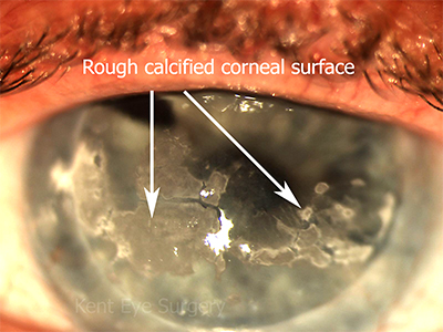
This is the deposition of calcium under the corneal epithelium. It tends to occur between the eyelids (in the interpalpebral zone). It starts just internal to the limbus, leaving a clear zone between the limbus and the affected area of the cornea and progresses across the visual axis. Initially, it is smooth and doesn’t disrupt the overlying corneal epithelium, but later it builds up to such an extent that the corneal epithelium is breached, when the condition becomes painful. The calcium can be removed surgically, which is very effective for rough plaques of calcium. Smooth band keratopathy is best removed with excimer laser (phototherapeutic keratectomy), although this is likely to cause a hypermetropic shift in refraction (it makes you more long-sighted).
Mild (smooth) band keratopathy is most commonly seen in patients with chronic glaucoma. More extensive cases, likely that illustrated above, is typically seen with chronic corneal exposure or neurotrophic keratopathy, particularly in patients with chronic herpes zoster keratitis.


