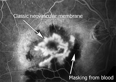
Predominantly classic (20%)
This is an example of a well defined “classic” membrane under the fovea in a patient with CSR. You should see the white area lightening up in the middle (fovea) of the central black area known as the macula. The other white spots represent malfunction of the retinal pigment epithelium in other areas of the retina in this condition.
Minimally classic (15%)
In these patients, the fluorescein angiogram is predominatly occult in picture, but small areas of well-defined CNV may be observed.
Occult (65%)
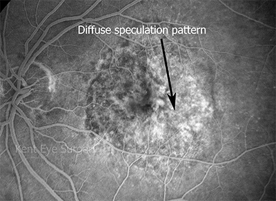
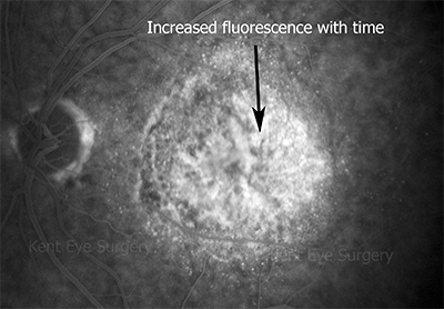
This is an example of occult CNV. There isn’t a well defined net as in the above example. The fluorescence leakage increses from early pictures:-


